
Extreme Lateral Interbody Fusion (XLIF)
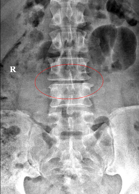
XLIF or Extreme Lateral Interbody Fusion surgery is a type of spinal fusion performed by which the surgeon accesses the intervertebral disc space from the side, lateral approach. This is in contrast to the more conventional anterior (from the front), and posterior (from the back) approach. Similar to any other interbody fusion surgery, the goal of the incision is to reach and replace the damaged disc, with a spacer device, fuse the opposing vertebrae.
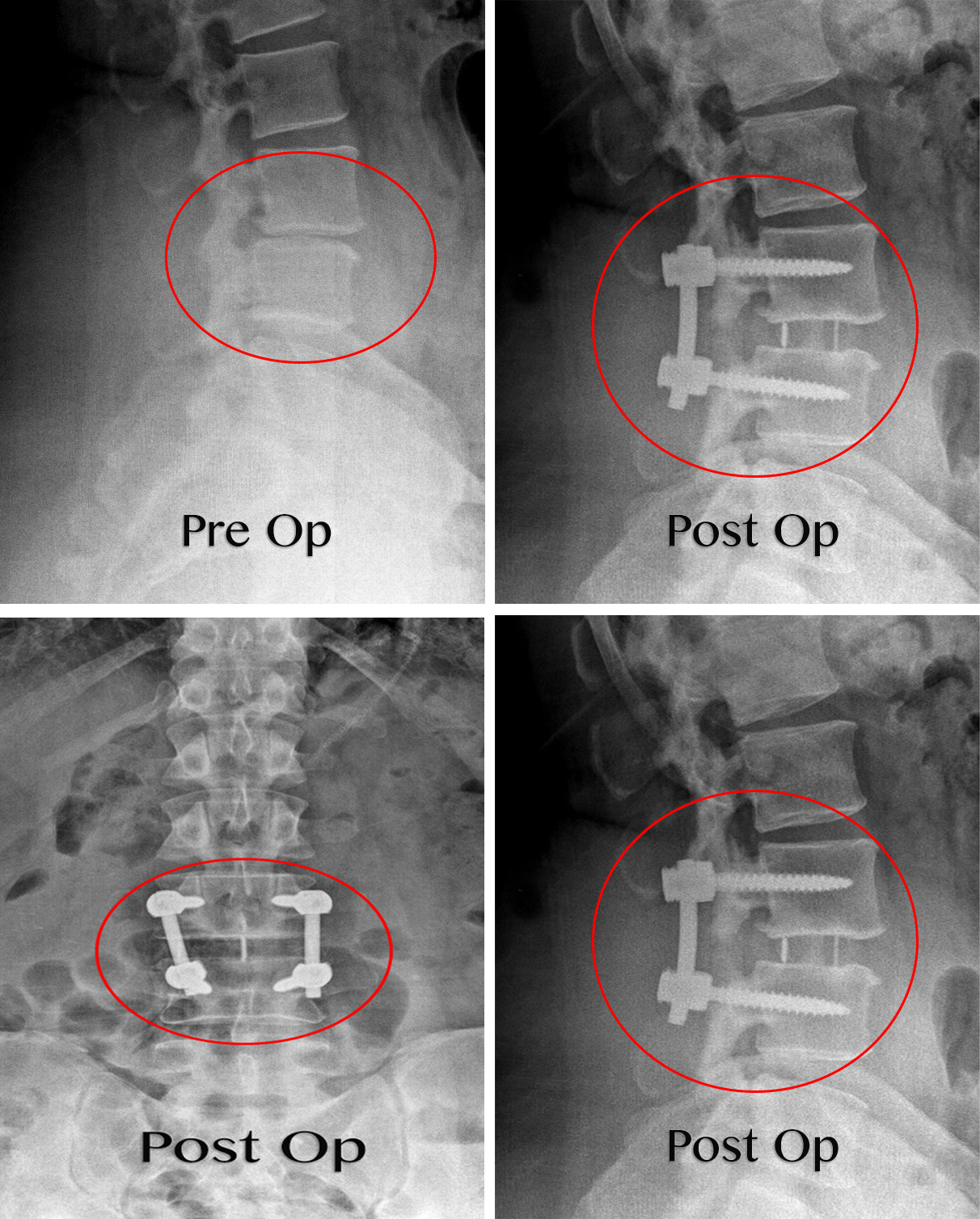
Benefits of XLIF
Some other common reasons why XLIF would be chosen over other procedures are:
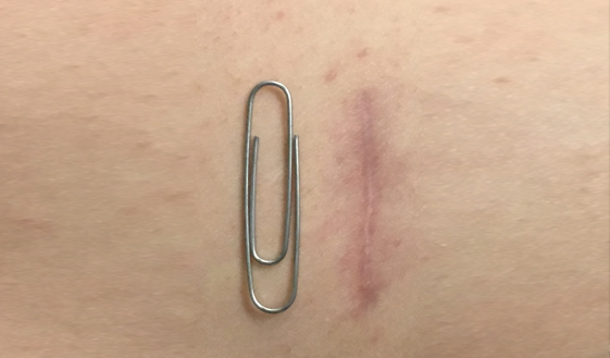
Why XLIF?
Those patients with certain issues affecting the lumbar spine or lower back are great candidates for XLIF. Some of the common conditions that can be addressed using XLIF are:
- Spinal Stenosis
- Degenerative Disc Disease
- Spondylolisthesis
- Scoliosis
- Other issues affecting the Lumbar Spine.
The XLIF is one of a number of spinal fusion options that a surgeon may recommend to treat specific types of lumbar spinal disorders, it is a similar procedure to ALIF, or PLIF, simply using a different approach. There are a number of reasons why your surgeon would choose to use a lateral (from the side) approach, as opposed to an anterior or posterior. Generally, it would be because the damaged disc, in need of replacement, is easier to reach through a lateral incision, therefore making for a far more efficient procedure, with less likelihood for complications.
How Does XLIF Work?
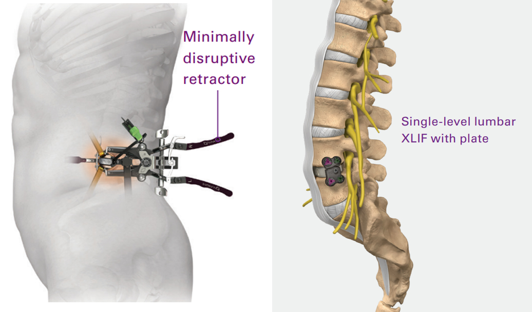
The procedure begins with the patient lying on his or her side. Using an x-ray or other methods, we locate the disc which needs to be removed. Once located, the surgeon marks the skin with a marker directly above the disc, and creates a small incision in the flank area, pushing away the peritoneum (a sac covering the abdominal organs from the abdominal wall.
The surgeon will then make a second incision directly on the patient’s side, and insert a tube-like device known as a dilator. This is helpful as the proximity to certain nerve roots, means the surgeon must have a real-time understanding of where the organs and nerve are located, relative to all instruments. Neuromonitoring is also done, a process which checks on the function of the nerves during surgery, to make sure the procedure stays on track.
The surgeon will usually have to split a muscle that is blocking access to the disc, known as the psoas muscle. Once access to the disc is made, it is removed, and a spacer is put into place. The body will naturally fill the spacer with bone, and the surrounding vertebrae will fuse together. Depending upon the individual needs of the patient, additional plates, screws and more may be added. If you are suffering from a degenerative disc issue or any pain in the lower back, you may be in need of interbody fusion surgery, contact Dr. Vikas Varma today and find out.