
Minimally Invasive Microdecompression
Minimally Invasive Microdecompression is done similar to the microdiscectomy as it relieves the compression of the nerve roots in the lumbar spine, by removing overgrown vertebral bone and soft tissue from the spine.
Benefits of MIS Microdecompression
Because the procedure is minimally invasive, there are a number of benefits that minimize the side effects. The procedure only requires a set of small incisions be made as opposed to one larger incision. The most notable advantages are:
Symptoms
Nerve compression will generally cause pain, numbness, or weakness in the arms, back, foot, or leg. Patients may also experience muscle issues, such as spasms, or overreactive reflexes, and even an uncomfortable tingling or burning feeling. The nerve issue can be caused by a number of spinal issues such as a herniated disc, or spinal stenosis.
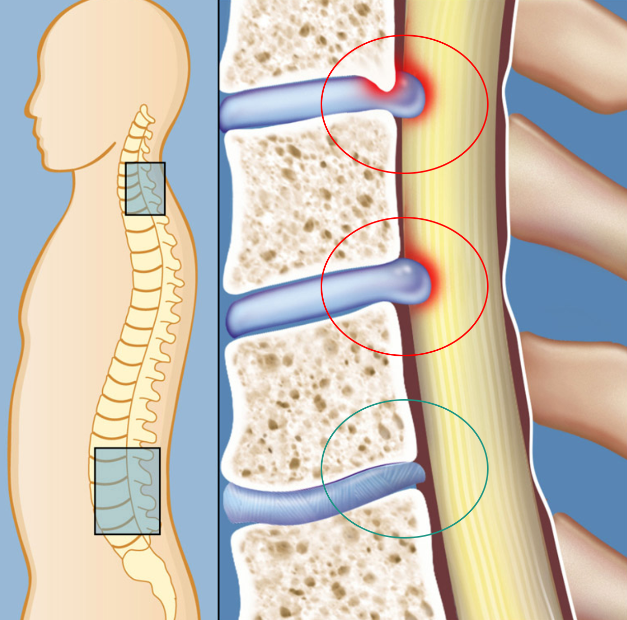
The red circles show points of Spinal Stenosis, at which the spinal canal has narrowed as a result of disc issues. This compression will require some form of decompression. The green circle, shows the normal disc, without any narrowing.
How Does MIS Microdecompression Work?
Using an X-ray the surgeon will identify the damaged/diseased discs or vertebral levels, and will create a small incisions directly above the site of the issue. Using a series of dilators, the surgeon will dilate the muscles that are preventing access to the spinal cord, and using a retractor creates a small channel between the muscles, whereby the surgeon can access the vertebra, as well as the compressed nerve root. Using the retractor minimizes any muscular damage as well as any pain you may feel. An endoscopic device may be inserted to provide a camera guide for the surgeon to perform the procedure through.
Any excess bone or ligament tissue is carefully removed, from the space around the nerve root helping to relieve any pressure or pain on the nerve. By repositioning the retractor and pointing the channel to the other vertebra, the surgeon can remove any other excess bone or tissue that may be causing further compression to the spinal nerve. The compression to the spinal nerve root has been relieved. All instrumentation is removed, and the incision is closed.
Patients can generally go home immediately following the procedure, however, some may need to stay for further monitoring. Since the procedure requires little to no downtime, patients are able to perform light activity almost immediately and will be able to resume their normal activities within 1-4 weeks of the procedure, depending on their body and the level of correction needed.
If you are suffering from nerve root compression resulting in pain in your arms, legs, or back, contact Dr. Vikas Varma, and schedule your appointment for treatment today.